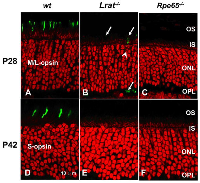
Figure 4. Confocal immunolocalization of cone opsins in wt, Lrat−/− and Rpe65−/− retinae.
(A–C) P28 wt and mutant retinae are stained for M/L-opsin. The M/L-opsin is nearly absent at this stage. A perinuclear ring (B; arrowhead) represents the endoplasmic reticulum-region where M/L-opsin (arrow) is synthesized. (D–F) P42 wt and mutant retinae are stained for S-opsin. The S-opsin is completely missing in the Lrat−/− and Rpe65−/− retinae at this stage, whereas the wt cones appear healthy with the opsin in the outer segments. Nuclei are contrasted with propidium iodide (red); scale bar represents 10 μm. All retinae sections passed through the optic nerve, and photoreceptors were imaged ventral (inferior) to the nerve where the degeneration was most advanced. Abbreviations as in Figure 1A.

