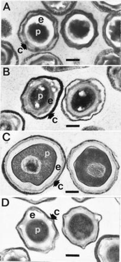
Figure 2.

Transmission electron microscopy of dormant and germinated wild-type and cwlD mutant spores. Dormant spores or spores that had been exposed to germinants for 60 min at 37°C were fixed with glutaraldehyde, postfixed, stained, and photographed as described (20). (A and B) Wild-type and cwlD mutant dormant spores, respectively. (C and D) Wild-type and cwlD mutant germinated spores, respectively. c, spore coats and exosporium; e, spore cortex peptidoglycan; p, spore protoplast. (Bar = 0.25 μm.)
