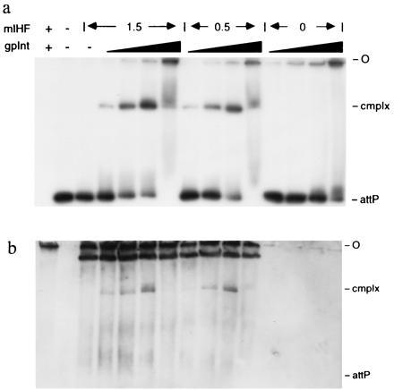
Figure 5.

Presence of mIHF in Int–mIHF–attP DNA complexes. Complexes were formed by addition of Int and mIHF to attP DNA, and the complexes were analyzed by native gel electrophoresis. All reactions contained 1 μg salmon sperm DNA and with the exception of the leftmost lane, contained ≈0.5 μg EcoRI–BamHI digested pCPΔR11 DNA of which the 364-bp attP fragment was radiolabeled. The three sets of reactions shown contain either 1.5 μM, 0.5 μM or no mIHF as indicated, and increasing amounts of Int (14, 48, 144, and 480 nM). The leftmost lane (no DNA control) contained 0.5 μM mIHF and 144 nM Int. An autoradiograph of the wet gel immediately after electrophoresis is shown in a, and an immunoblot of the same gel with anti-mIHF serum is shown in b. The origin of electrophoresis (O) and the positions of attP DNA and Int–mIHF–DNA complex (cmplx) are shown. Other bands in the immunoblot probably correspond to mIHF nonspecifically bound to DNA. The reactions in the absence of mIHF (last four lanes) show that the antiserum does not cross-react with Int.
