
Figure 1.
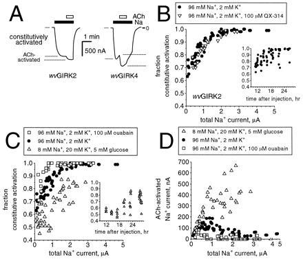
Constitutive activation of wvGIRK2 and wvGIRK4 is increased by intracellular Na+. (A) Representative traces from oocytes injected with 3 ng m2AChR mRNA plus either 5 ng wvGIRK2 mRNA or 25 ng wvGIRK4 mRNA, held at a membrane potential of −80 mV. The traces show the constitutively activated Na+ current and the acetylcholine (ACh)-activated Na+ current. Bars indicate application of 98 mM Na+ and 1 μM ACh; where no bar is shown, the bath solution contained 98 mM NMDG as the only monovalent cation. (B) Variation of constitutive activation with total Na+ current for wvGIRK2-injected oocytes incubated in the standard medium (96 mM Na+/2 mM K+) with (•) or without (▿) 100 μM QX-314, but tested in the absence of QX-314. Each data point represents a single oocyte from either three or two separate batches, respectively. (Inset) Variation of constitutive activation with time since injection. (C) Variation of constitutive activation with total Na+ current for wvGIRK2-injected oocytes incubated in the standard medium alone (•), standard medium with 100 μM ouabain (□), or 8 mM Na+, 20 mM K+ with 5 mM glucose (▵). Each data point represents a single oocyte from either two, three, or one separate batch(es), respectively. (Inset) Variation of constitutive activation with time since injection. (D) The data of C are presented as ACh-induced currents for the three experimental manipulations.