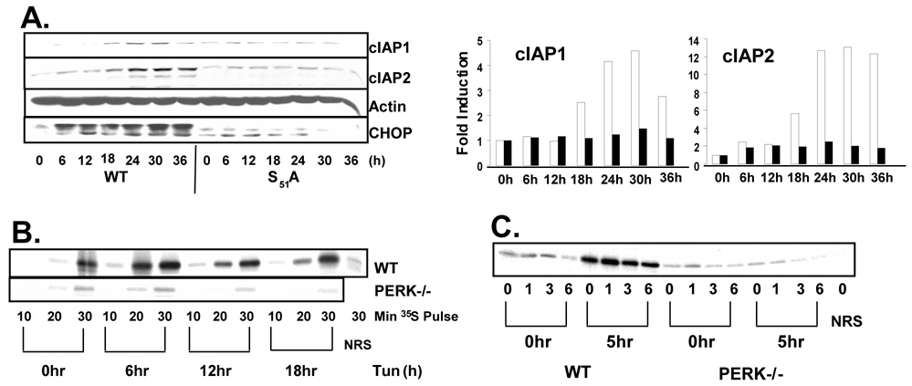
Figure 5. IAP mRNA is selectively translated during ER stress.
(A) Wild-type and eIF2α S51A fibroblasts were treated with tunicamycin as indicated. Cell lysates were resolved by SDS-PAGE and IAP levels were determined by immunoblot. Graphs represent fold increase in cIAP protein. Wild-type (white) and S51A (black). (B) Wild-type and PERK−/− fibroblasts were treated with tunicamycin or left untreated. Cells were pulsed with 35S methionine during the last 10, 20, or 30 min of treatment and cIAP2 was immunoprecipitated from cell lysates and observed by autoradiograph. (C) Wild-type and PERK−/− fibroblasts were treated with tunicamycin for 5 hours or left untreated. Cells were then pulsed with 35S-mentionine for 30 min followed by chase in cold methionine for the indicated intervals. cIAP2 was immunoprecipitated from cell lysates and observed by autoradiograph. NRS lanes represent immunoprecipitation with Normal Rabbit Serum.

