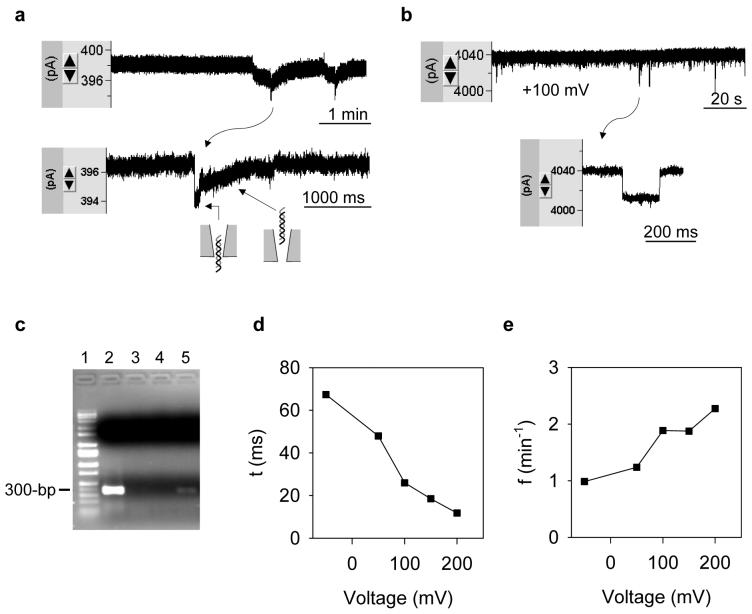
Figure 3.
Translocation of dsDNA in nanopores. a. Current blocks by 1 kb DNA (10 nM) in the external solution (1 M NaCl) through a 3.8 nS nanopore (+100 mV). b. Current blocks with DNA through a 39 nS nanopore (+100 mV). c. Reveals DNA translocation by PCR. Lane-1, marker; Lane-2, 10 μl bath solution (containing DNA); Lane-3, 10 μl dd-H2O; Lane-4, 10 μl internal solution without DNA (control); Lane-5, 10 μl internal solution near nanopore after the DNA translocation. d. Voltage-dependent duration of DNA translocation. e. Voltage-dependent occurrence of DNA translocation.

