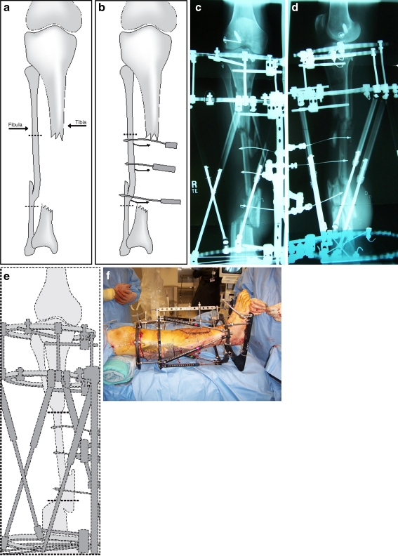
Fig. 2.
a This is an illustration of the 15-cm tibial defect. b A diagrammatic representation of the fibular transport shows the level of the fibular osteotomy and the direction of the fibular transport using three olive fibular pulling wires. c, d The AP/lateral X-ray views of the leg were taken after the application of the two-ring frame and the fibular osteotomies. Note the three fibular wires aiming from posterolateral to anteromedial direction. e This is a diagrammatic representation of the fibular transport in the Ilizarov/TSF with three pulling olive wires. f This picture, taken in the operating room, is of the patient's leg after the application of Ilizarov/TSF2

