


Figure 3. Vesicular Sepp1 staining observed in all Apoer2 ICD mutant mice.
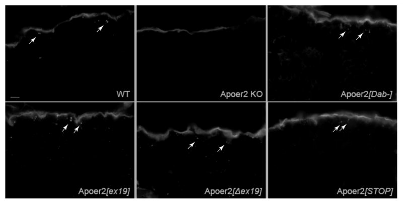
Fixed mouse testes were stained for Sepp1. Vesicular Sepp1 staining (arrows) is apparent in WT mice and all Apoer2 ICD mutants. As previously reported, there is no Sepp1 vesicular staining in Apoer2 KO testes. Scale bar = 10 μm.