


Figure 4. Sperm immunostaining reveals normal organization of spermatozoa in all Apoer2 mutant mice.
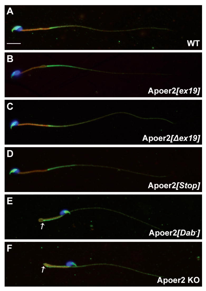
Fixed spermatozoa were immunostained with MitoFluor™ (midpiece; red), a crossreacting polyclonal antibody against an unidentified sperm antigen (acrosome and principal piece; green), and DAPI (nucleus; blue). (A) wild type (B) Apoer2[ex19] (C) Apoer2[Δex19] (D) Apoer2[Stop] (E) Apoer2[Dab-] (F) Apoer2 KO. Pictures are representative of the major morphological sperm population for each genotype. Arrows indicate location of annulus. Scale bar = 10 μm.