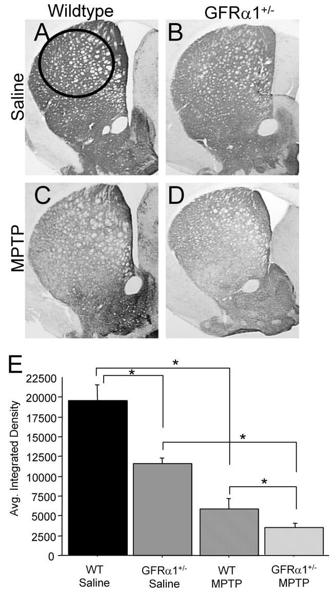
Figure 4. GFR α1+/− mice treated with MPTP demonstrated a greater loss of striatal tyrosine hydroxylase (TH) immunoreactivity (ir) than WT mice.
Photomicrographs of TH-ir in coronal hemisections of the striatum from (A) saline-treated WT mice, (B) saline-treated GFR α1+/−, (C) MPTP-treated WT mice, and (D) MPTP-treated GFR α1+/−. (E) Quantification of the average integrated density using one-way ANOVA analysis followed by Student-Nuemann-Kuels post-hoc test from the four treatment groups at 26 months of age confirmed that there were significant differences in the striatum between GFR α1+/− and WT mice treated with saline (*p<0.05). The administration of MPTP resulted in a significant reduction of striatal TH-ir compared to saline-treated mice, regardless of genotype (*p<0.05), with the greatest loss of TH-ir in the striatum of GFR α1+/− mice. Photomicrograph magnification=2x. N=8 per group. Error bars indicate SEM.

