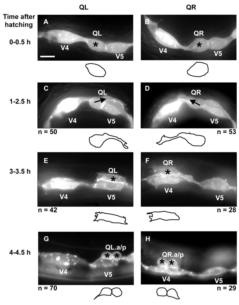
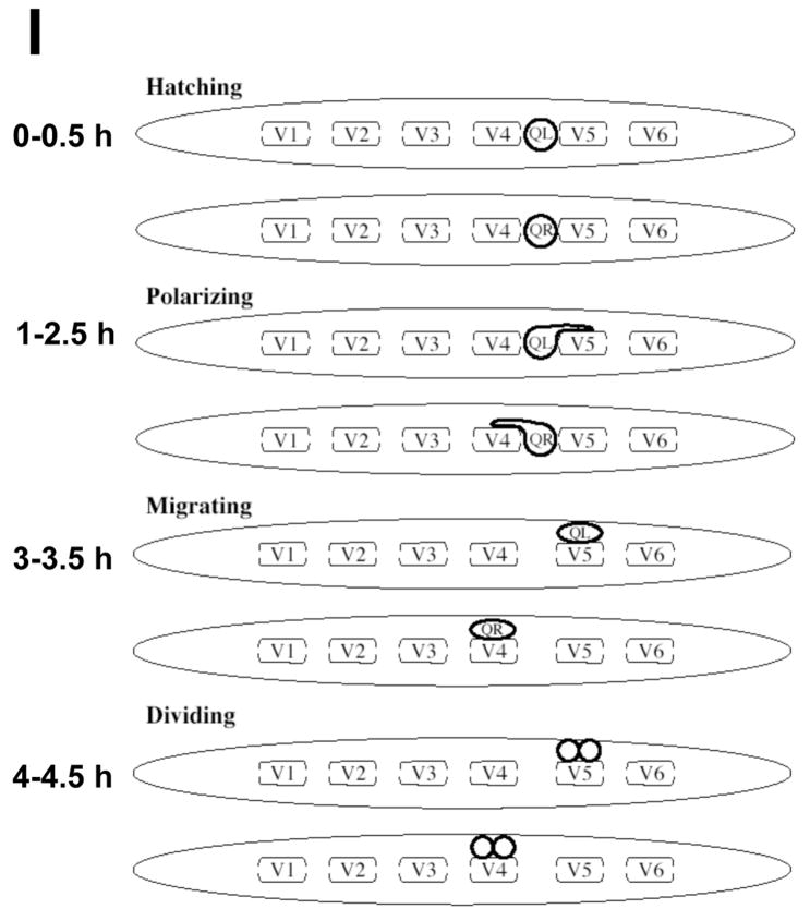
Figure 2. Q neuroblast polarization and migration.
(A–H) are panels of epifluorescent micrographs of Q neuroblasts of L1 larvae at given timepoints after hatching expressing the scm::gfp::caax marker in the Q cells and the lateral seam cells (V cells). Tracings of the boundaries of the Q neuroblasts in each micrograph are located below each panel. For each time point, the QL neuroblast is found on the left (A,C,E,G) and the QR neuroblast is found on the right (B,D,F,H). The scale bar in (A) represents 5μm for (A–H). (A–B) Unpolarized Q neuroblasts visualized 0–0.5 h after hatching. Asterisks mark the position of the Q neuroblasts. (C–D) Polarization of the Q neuroblasts visualized 1–2.5 h after hatching. QL sent a process posteriorly over the V5L seam cell. QR sent a process anteriorly over the V4R seam cell. Arrows indicate the direction of polarization of the Q neuroblasts. (E–F) Migration of the Q neuroblasts visualized 3–3.5 h after hatching. QL migrated posteriorly over the V5L seam cell. QR migrated anteriorly over the V4R seam cell. (G–H) Division of the Q neuroblasts visualized 4–4.5 h after hatching. QL divided over the V5L seam cell to produce QL.a and QL.p. QR divided over the V4R seam cell to produce QR.a and QR.p. Asterisks mark the position of the Q neuroblast daughters. (I) A schematic diagram demonstrating the polarization, migration, and division patterns of the Q neuroblasts.


