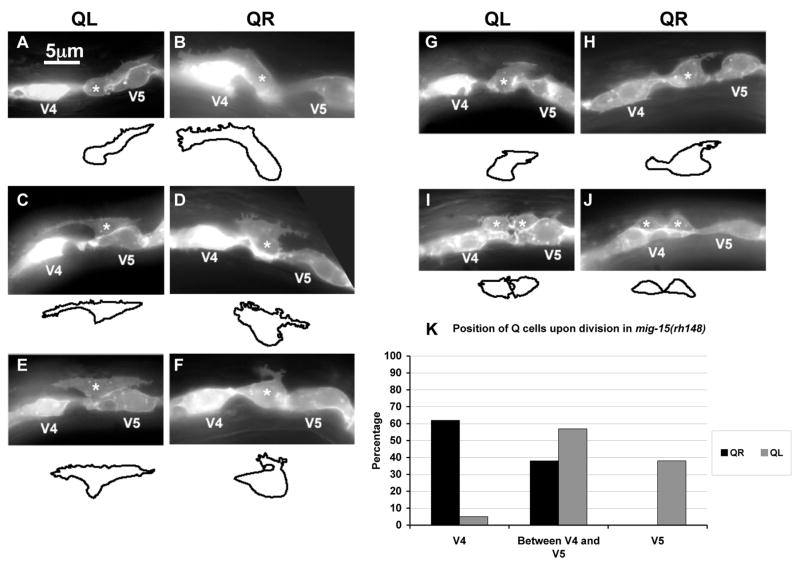
Figure 3. mig-15 NIK kinase hypomorphic mutation affects the polarizations and migrations of the Q neuroblasts.
(A–J) are panels of epifluorescent micrographs of Q neuroblasts of L1 larvae in mig-15(rh148) mutants with scm::gfp::caax expression. Asterisks mark the position of the Q neuroblast at 3–3.5 h after hatching (A–H) or Q neuroblast descendants at 4–4.5 h after hatching (I–J). Tracings of the Q neuroblasts found in each micrograph are located below each panel. The scale bar in (A) represents 5μm for (A–J). (A) A QL neuroblast polarized normally over the V5L seam cell. (B) A QR neuroblast polarized normally over the V4R seam cell. (C) A QL neuroblast failed to maintain proper posterior polarization, and sent a protrusion to the anterior. (D) A QR neuroblast failed to maintain proper anterior polarization, and sent a protrusion to the posterior. (E) A QL neuroblast sent protrusions to both the anterior and posterior. (F) A QR neuroblast was not strongly polarized in either direction and sent a small, anteriorly-directed protrusion from the posterior of the cell. (G) A QL neuroblast was not strongly polarized in either the anterior or posterior direction, although maintained slight posterior polarization. (H) A QR neuroblast was not strongly polarized in either the anterior or posterior direction and sent a small protrusion posteriorly. (I) A QL neuroblast divided between the V4L and V5L seam cells. (J) A QR neuroblast divided over the V4R seam but was not atop the V4R seam cell. (K) Quantitation of the position of the QL and QR neuroblasts upon division (4–4.5 h after hatching) in mig-15(rh148) mutants. Position with respect to the V4 and V5 seam cells is the X axis, and the percentage of Q neuroblast daughters found at those positions is the Y axis. For QL divisions, 37 animals were scored. For QR divisions, 26 animals were scored.

