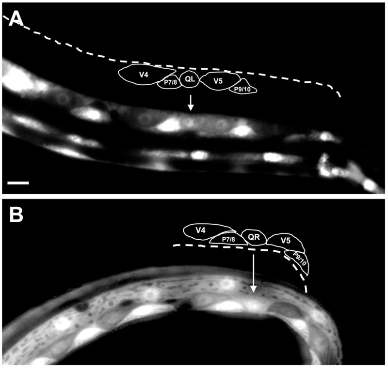
Figure 9. mig-15 is expressed in the Q neuroblasts.
Shown are epifluorescent micrographs of newly-hatched L1 larvae expressing the mig-15::gfp transcriptional promoter fusion. Tracings of the Q cells and the surrounding cells are shown above each micrograph. Arrows mark the positions of Q cells. (A) A left ventral-lateral view of an animal with mig-15::gfp expression in QL. The lateral seam cells (V cells) and P cells also express mig-15::gfp. (B) A right dorsal-lateral view of an animal with mig-15::gfp expression in QR. In this animal, the V5 seam cell lost mig-15::gfp and showed no fluorescence. The scale bar in (A) represents 5μm for (A–B).

