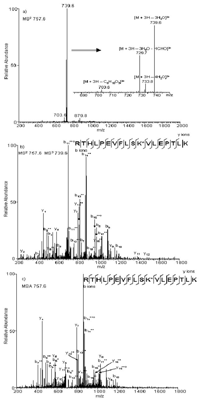
Figure 8. Comparison of spectra obtained under standard and advanced CID-MS/MS.
(a) Product-ion spectrum produced from CID-MS/MS fragmentation of the [M + 3H]3+ ion of peptide RTHLPEVFLSK*VLEPTLK, where * represents the Amadori modification site. Different neutral-losses are shown in the zoomed inset. Product-ion spectra produced from the [M + 3H]3+ ion of peptide RTHLPEVFLSK*VLEPTLK under b) NLMS3 triggered by neutral-loss of 3 H2O or c) MSA triggered by neutral-loss of 3 H2O, where * represents the Amadori adduct modification site.

