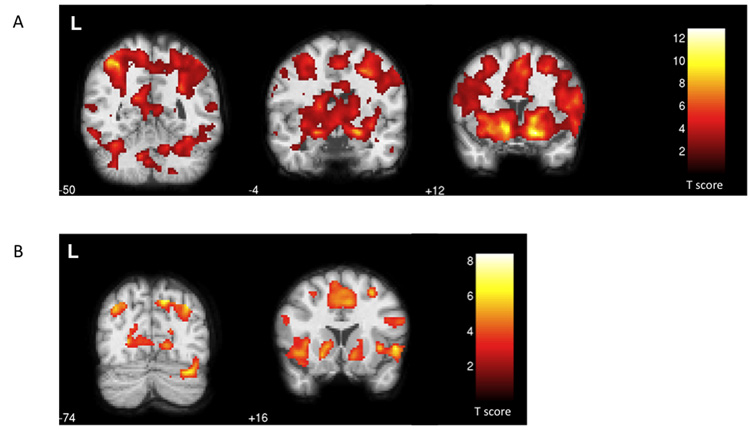
Figure 3.
Statistical parametric maps of the functional localization within the coronal plane (p <0.05, FDR-corrected). a) Regions responding to positive feedback > uninformative feedback included the bilateral cerebellum (z = −50), the bilateral amygdala (z =− 4), and the nucleus accumbens, caudate nucleus, putamen, insula, and SMA bilaterally (z = +12). b) For the negative feedback > uninformative feedback comparison, activated regions included the right cerebellum (z = −74) and the bilateral nucleus accumbens, caudate nucleus, putamen, insula, and SMA (z = +16).

