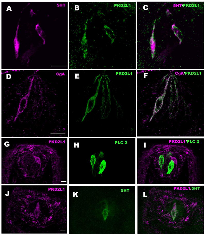
Figure 2.
LSCM images of double-labeled longitudinal sections of circumvallate taste buds labeled with markers for Type III taste cells: PGP-9.5 (A-C), NCAM (D-F), 5-HT (G-I) and CgA (J-L) along with PKD2L1. A-C: PKD2L1-IR is present in the PGP-9.5-IR taste bud cells. However not all cells positive for PGP-9.5 showed PKD2L1-IR (arrow), corresponding to the presence of PGP-9.5 in some Type II cells (Yee et al. 2003). D-L: PKD2L1-IR colocalizes with markers of Type III cells: D-F: NCAM immunoreactive nerve fibers appear as punctate staining between the larger, immunoreactive taste cell profiles. G-I: 5-HT and J-L: CgA. Scale bars = 20 μm.

