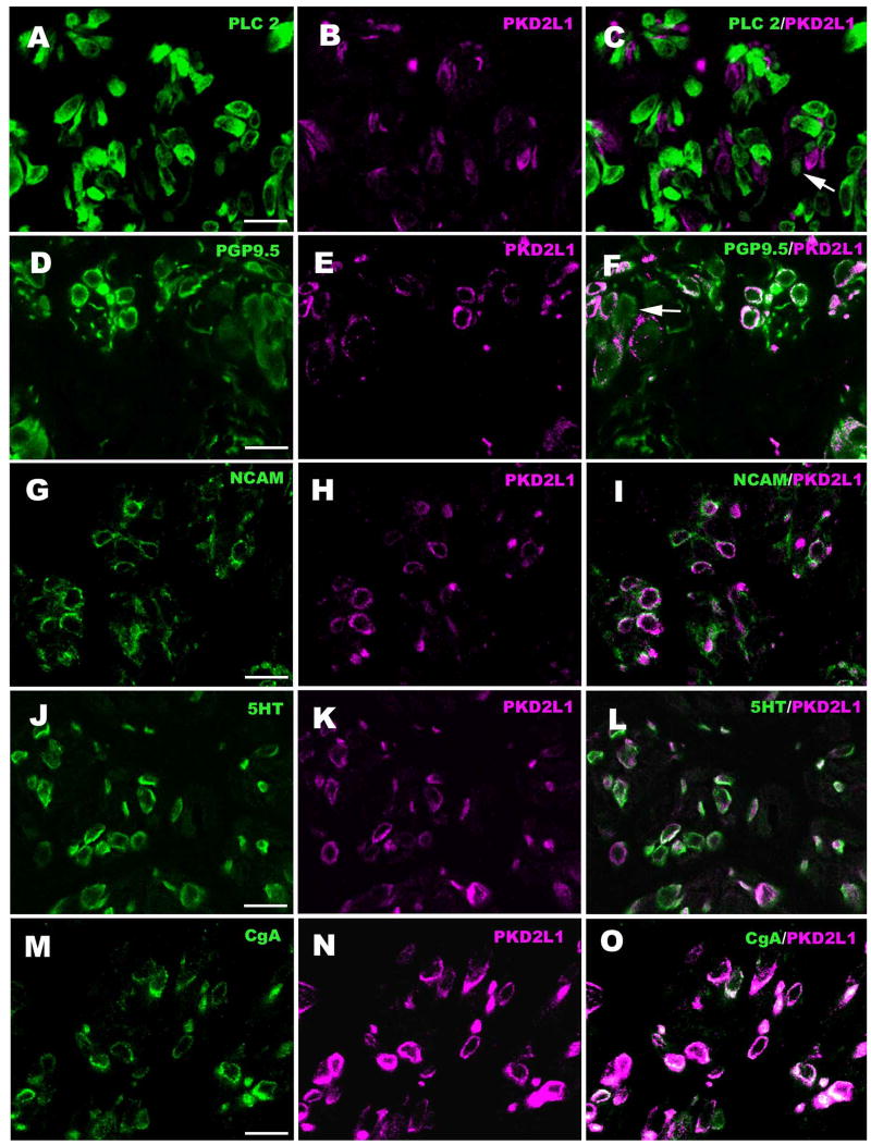
Figure 3.
Double-labeled cross-sections through the upper part of circumvallate taste buds showing lack of co-localization of PLCβ2 (Type II cell marker) and PKD2L1, but substantial co-localization with PGP-9.5 which reacts with both Type II and Type III taste cells. Some PGP-9.5-IR cells do not express PKD2L1 (arrow, panel F). The Type III cell markers, NCAM, 5-HT, and CgA essentially completely co-localize with PKD2L1. Green indicates PLCβ2-IR (A), PGP-9.5-IR (D), NCAM-IR (G), 5-HT-IR (J) and CgA-IR (M), and magenta indicates PKD2L1-IR (B, E, H, K, N). Scale bars = 20 μm.

