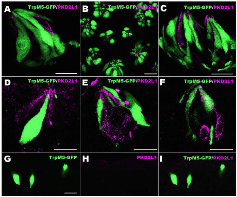
Figure 4.
Dual label images showing PKD2L1 (magenta) and GFP as expressed in TrpM5-GFP mice. A-C: These LSCM overlay images shows that taste cells positive for PKD2L1 do not express TrpM5 in longitudinal (A) and cross (B) sections of circumvallate, and foliate (C) taste buds. D-F: Expression of PKD2L1 in non-lingual taste buds. These images show that PKD2L1 is present in palate (D), pharynx (E) and larynx (F) taste buds. PKD2L1-expressing taste cells are different from those expressing TrpM5. G-I: Laryngeal solitary chemoreceptor cells (SCCs) are shown (green) in TrpM5-GFP mice. PKD2L1-IR is not detectable in these SCCs of the larynx. Scale bars = 20 μm.

