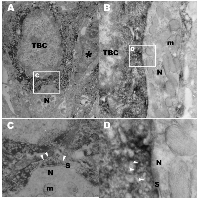
Figure 5.
DAB immuno-electron micrograph of taste buds in the circumvallate papilla showing PKD2L1-IR taste bud cells (TBC) with two structural types of synapses onto sensory nerve processes. Synapses between taste cells and nerve processes are a characteristic of Type III taste cells. A: A Synapse onto a nerve process (N) indented into the soma of the taste cell is associated with PKD2L1-IR taste bud cell (TBC). An adjacent taste bud cell (*) lacks PKD2L1-IR. B: A macular synapse from a taste bud cell (TBC) with PKD2L1-IR onto a nerve process (N). Numerous mitochondria (m) are present in the postsynaptic process. C: High magnification of boxed area in A showing synapse (S). Many synaptic vesicles (arrow heads) are evident in the cytoplasm opposite the point of contact with the nerve fiber. Some mitochondria (m) are also present in the postsynaptic region. D: High magnification of boxed area in B showing synapse (S). The PKD2L1-IR taste bud cell contains many clear synaptic vesicles (arrow heads) in the presynaptic region. Scale bars=1 μm in A, B; 0.25 μm in C, D.

