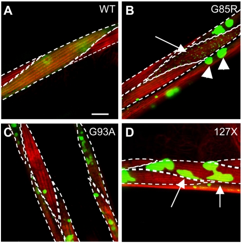
Figure 3. Mutant SOD1 forms morphologically distinct aggregates.
Projections of confocal Z-stacks with several adjacent muscle cells; punctate lines delineate individual cells, red staining shows myofilaments stained with Rhodamine-phalloidin. G85R protein (B) forms morphologically diverse aggregates, including large foci indicated by arrowheads and small dispersed foci, indicated by arrow. Aggregates in about 40% of cells of 127X animals appear as irregular, elongated foci (D, arrows and inserts in Figure 1 J–L ). Scale bar in A is 10 micrometers. Images are selected as representative of typical aggregation morphology among at least 100 imaged cells per genotype.

