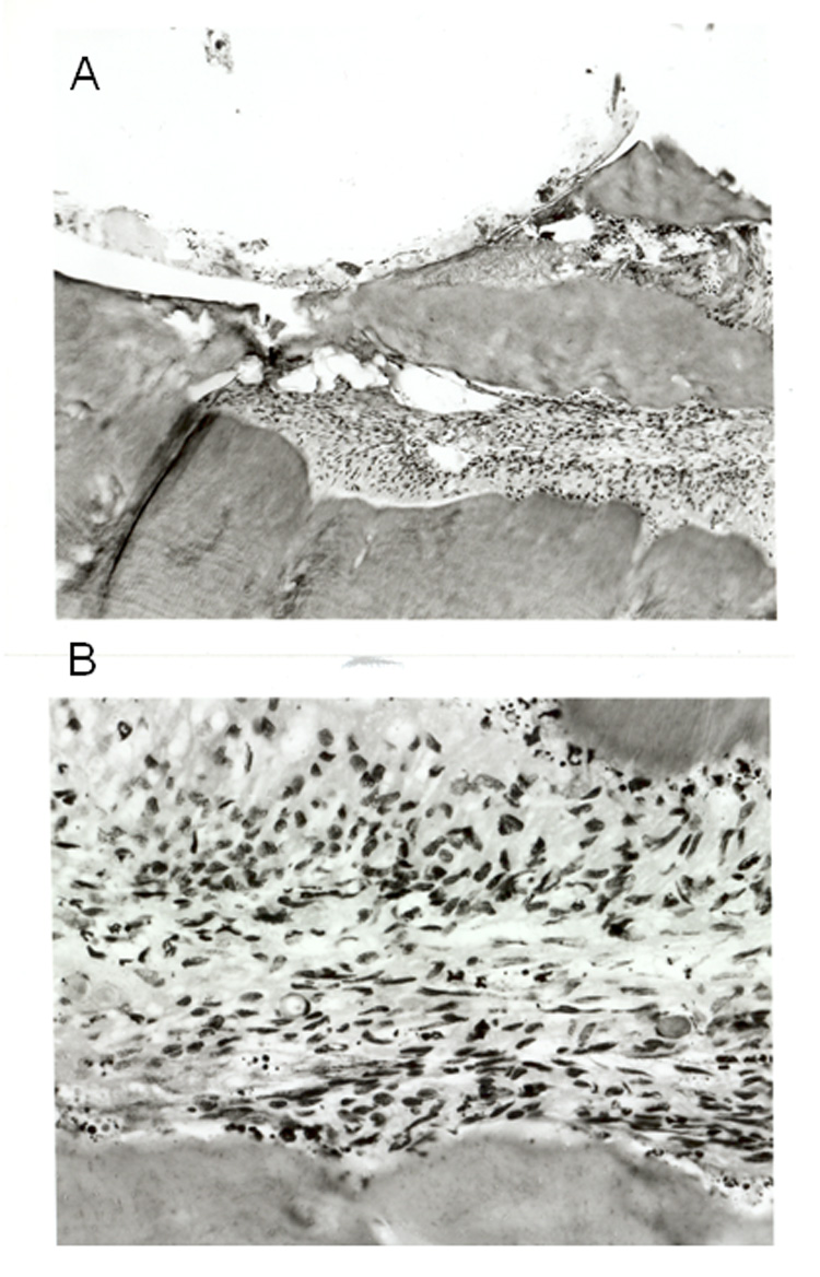
Figure 5.

Light microscopic photographs of hematoxylin-eosin stained sections taken from the manipulated teeth 48 hours following the procedure. A. A section (x10) taken from the drilled area (upper part of the picture) B. A section (x 40) taken from the pulp, note the numerous immune cells within the tissue.
