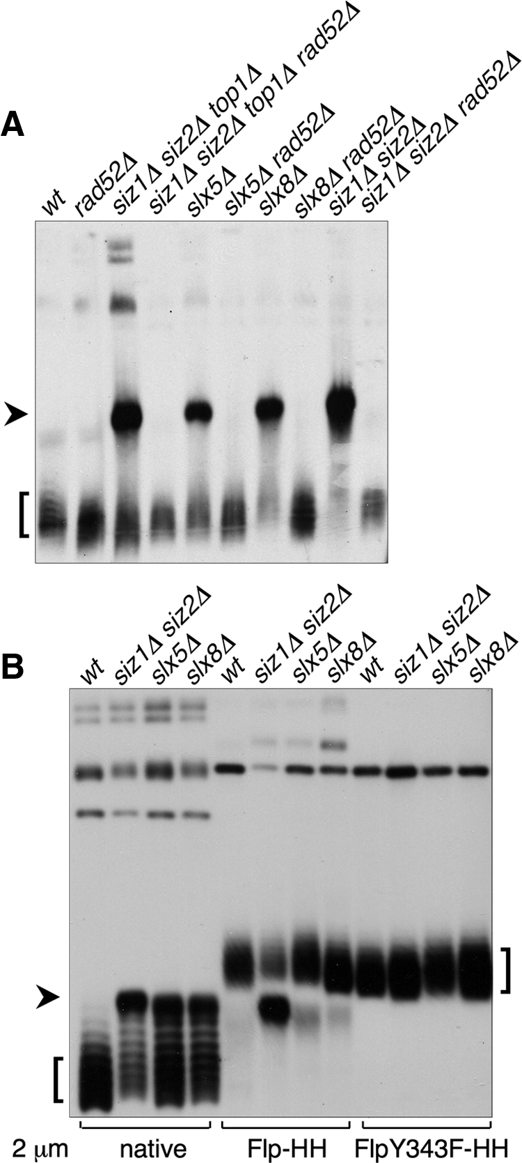
Figure 3.

Formation of a HMW 2 μm species in SUMO pathway mutants. (A) Uncut DNA from the indicated strains was analyzed by electrophoresis in an agarose gel containing chloroquine, followed by Southern blotting with a probe against 2 μm. Lanes were normalized to contain equal amounts of 2 μm DNA, as measured by qPCR. Arrowhead indicates the aberrant HMW species. Square bracket indicates supercoiled monomeric 2 μm. (B) DNA from strains of the indicated genotypes containing the indicated versions of 2 μm were analyzed as in A. Both the Flp-Y343F 2 μm variant and the corresponding wt control contained a marker gene, and consequently were larger than native 2 μm. Thus, the supercoiled monomer forms of these plasmids migrated more slowly than native 2 μm, but the HMW form ran at the same position as for native 2 μm.
