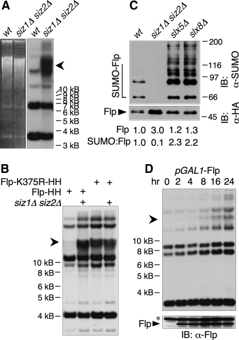
Figure 4.
Defects in Flp sumoylation lead to formation of the HMW species. (A) Uncut yeast DNA from the indicated strains was analyzed by EtBr-agarose electrophoresis (left) followed by Southern blotting with a probe against 2 μm (right). Lanes contained equal amounts of 2 μm DNA. An arrowhead indicates the HMW form. (B) Uncut DNA from wt or siz1Δ siz2Δ strains containing the indicated versions of 2 μm was analyzed by Southern blotting as in A. (C) Proteins from indicated strains containing 2 μm expressing Flp(Y343F)-HA-His8 (which does not allow 2 μm amplification) were purified by Ni-NTA affinity chromatography and analyzed by SDS-PAGE and immunoblotting with Abs against SUMO (top) and HA (bottom). Arrowhead indicates unmodified Flp, and an open bracket indicates SUMO-modified Flp. Levels of unmodified Flp as well as ratios of total SUMO signal to unmodified Flp are given below the lanes. Quantities are expressed relative to wt. (D) Flp was expressed from the galactose-inducible GAL1 promoter for the indicated times in log phase wt cells containing native 2 μm. A600 was kept ≤2.0. (top) Uncut DNA was analyzed by Southern blotting as in A, except that lanes contained equal amounts of total DNA rather than equal amounts of 2 μm DNA. (bottom) Whole cell lysates from the same samples as in top panel were analyzed by immunoblotting with an Ab against Flp. Asterisk designates a band that cross-reacts with the Ab.

