Figure 5.

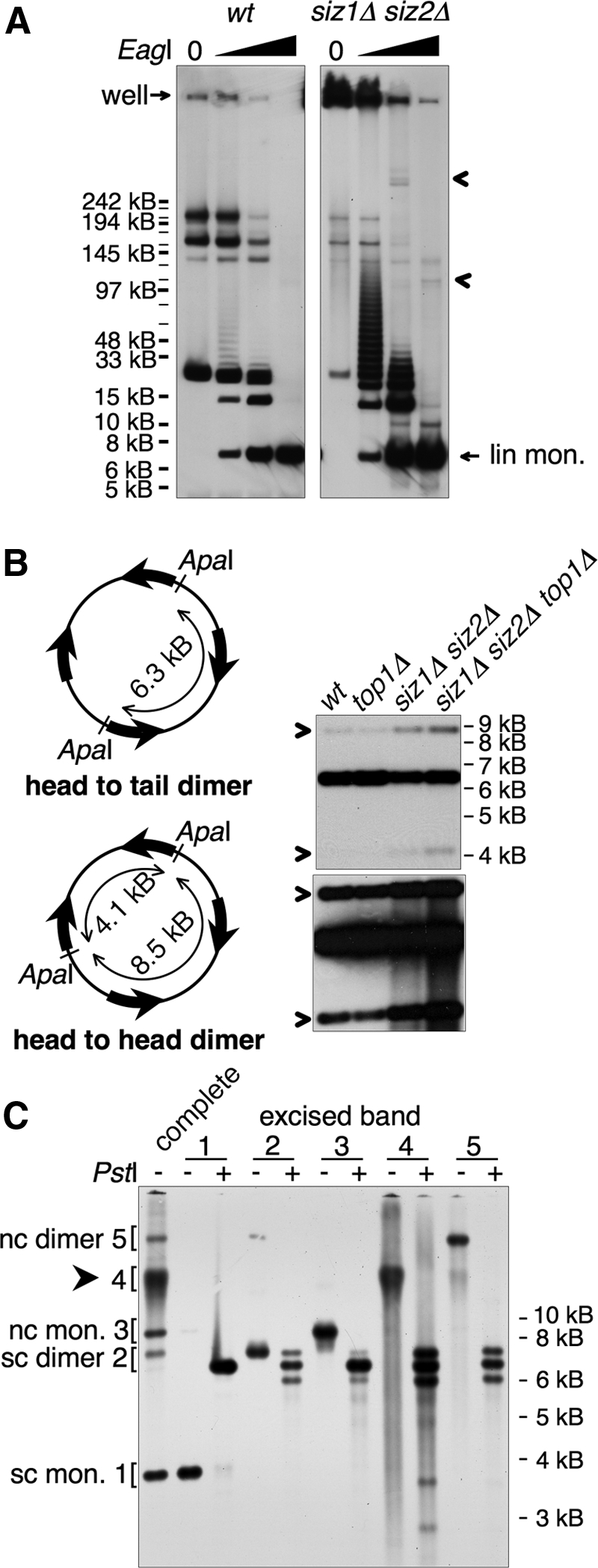
The HMW form contains tandem copies of 2 μm. (A) DNA from indicated strains was left untreated or cleaved with increasing amounts of EagI, as indicated, and analyzed by pulsed-field gel electrophoresis, followed by Southern blotting using a probe against 2 μm. All samples were cleaved to completion with BglII, which does not cleave in 2 μm, to reduce chromosomal DNA to small fragments. wt and siz1Δ siz2Δ panels are from different exposures from the same blot, chosen to normalize the signal from the fully cleaved 2 μm band to the same level, as quantified using the Chemidoc system. The fully cleaved 6.3-kb linear monomer and the position of the well are indicated. Arrowheads designate siz1Δ siz2Δ-specific low-mobility species released by partial restriction digest that may represent nonlinear portions of the HMW species. Markers were NEB MidRange I, which are all indicated, but not all labeled. Major bands in wt uncut sample probably represent the small circular forms of 2 μm, although this has not been shown directly. (B) Left, illustration of head-to-head and head-to-tail dimer linkages and ApaI restriction fragments. Wide arrows denote the 599-base pair internal repeat sequences that contain FRT. Right, DNA from the indicated strains was digested with ApaI and analyzed by Southern blotting with a probe against 2 μm. Bottom, a darker exposure of the top panel. Bands derived from head-to-head dimer linkages are designated with arrows. (C) The five bands indicated on the left were isolated from an EtBr-stained agarose gel and were either analyzed uncut or were digested with PstI, as indicated, followed by Southern analysis as in Figure 4B. Identities of the 2 μm species are indicated. sc, supercoiled; nc, nicked circular; mon., monomer. Arrowhead designates HMW species.
