Figure 5.
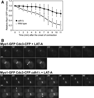
APCCdh1-dependent disassembly of the actomyosin ring during contraction (A) or in its absence (B). Strains YEF1681 (MYO1-GFP) and RNY2349 (MYO1-GFP cdh1Δ) were used. (A) Cells were examined by 4D time-lapse microscopy (see Materials and Methods). The intensities of Myo1-GFP, relative to the intensity measured at the beginning of ring contraction, are plotted for the indicated times (mean ± SD; n = 7 for each strain). All Myo1 rings had completed contraction by the final time point. No corrections were made for possible photobleaching during the course of the experiment; thus, the removal of Myo1 from the ring may have been even more defective in cdh1Δ cells than these data suggest. (B) Transformants containing plasmid YCp111-CDC3-CFP were treated with 200 μM LAT-A at time zero and examined by time-lapse microscopy at 1-min intervals. Top panels, Myo1-GFP; bottom panels, Cdc3-CFP. Arrowheads show the splitting of the septin hourglass structure into two rings, indicating the onset of cytokinesis. Representative images are shown. Bars, 2 μm.

