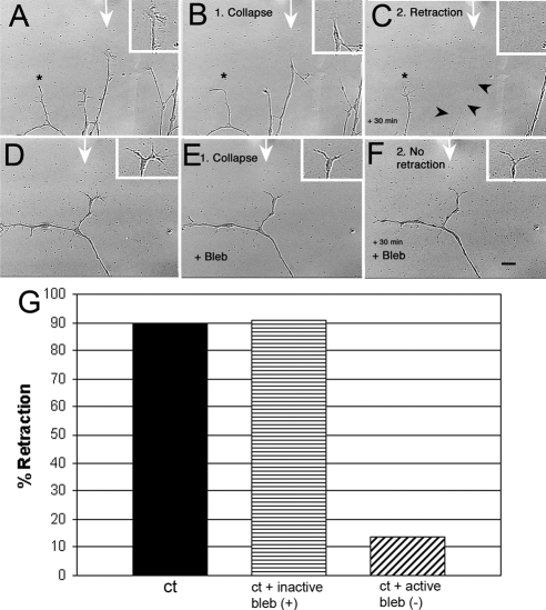
Figure 2.
DRG neurons for control mice show myosin II–dependent retraction in response to a Sema 3A gradient. Cultures were preincubated with either 100 μm inactive blebbistatin (+) or 10 μm active blebbistatin (−) for 30 min. A gradient of Sema 3A (7.5 μg/ml in the pipette) was applied for 30 min during time-lapse imaging (1-min intervals) via micropipette. The time-lapse movie was used to score for extension, collapse, retraction, and turning. (A–C) A sequence showing typical DRG neurite responses to a Sema 3A gradient when cells are grown on low laminin-1. The micropipette tip was just outside the field at the point indicated by the white arrow. A growth cone in line with the Sema 3A gradient first collapses (15 min) (B) and then retracts (30 min; C). Arrowheads, residual retracted neurite. An adjacent neurite (*) further from the gradient does not retract. (D–F) Blebbistatin (Bleb) treatment does not prevent collapse (E), but does prevent retraction (F). Scale bar, 10 μm. (G) Inactive blebbistatin (+) had no effect on retraction compared with untreated controls, but active blebbistatin (−) caused a significant drop in the number of cells retracting (p = 0.0001; Ct, n = 28; Bleb (−), n = 37; Bleb inactive, n = 11) indicating that myosin II activity is necessary for retraction in response to Sema 3A gradients.

