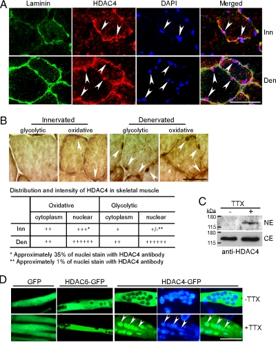
Figure 6.
Muscle inactivity stimulates nuclear accumulation of HDAC4. (A) Triple fluorescence immunostaining for laminin, HDAC4, and nuclei on innervated and denervated TA muscle sections. Arrowheads point to HDAC4+ myonuclei located within the muscle fibers basal lamina; 600× magnification. (B) HDAC4 immunoreactivity shows low-level nuclear accumulation of HDAC4 in innervated small oxidative fibers and high level nuclear accumulation in denervated small oxidative and larger glycolytic fibers. Arrowheads point to nuclei with weak HDAC4 immunoreactivity in oxidative fibers of innervated muscle and high HDAC4 immunoreactivity in both oxidative and glycolytic denervated muscle fibers; 1000× magnification. Qualitative assessment of HDAC4 expression in cytoplasm and nuclei is presented in the table. (C) Western blot analysis of HDAC4 nuclear levels (NE) and cytoplasmic levels (CE) in active (−TTX) and inactive (+TTX) primary mouse myotube cultures. (D) Inactivity induced nuclear accumulation of HDAC4 in cultured primary mouse myotubes. Primary myotubes were transfected with indicated expression vectors and either kept inactive with TTX or allowed to spontaneously contract (−TTX); 400× magnification. Bars, 40 μm. Inn, innervated; Den, denervated.

