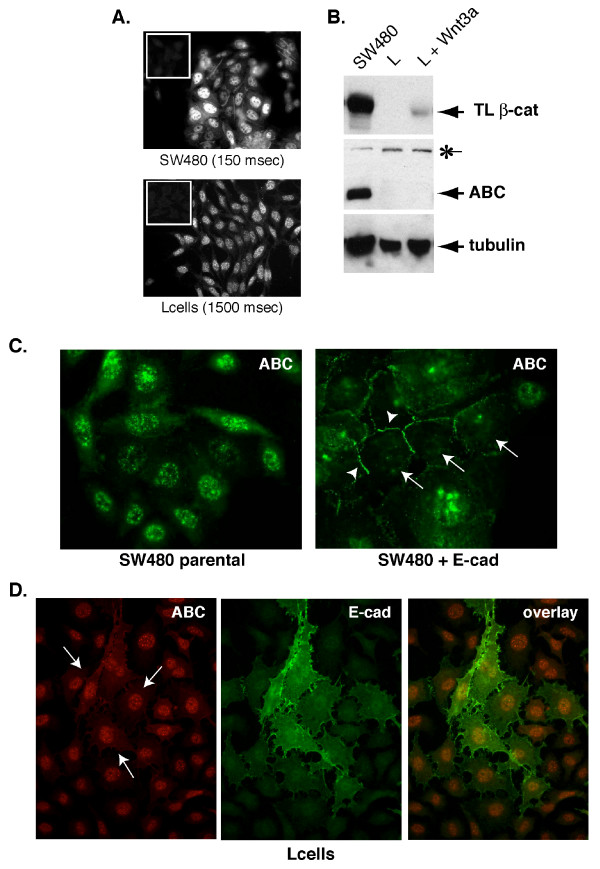
Figure 3.
Assessment of nuclear localized ABC can be verified by cadherin-mediated depletion of the nuclear signal. A&B. A. Immunofluorescence staining of ABC with mAb 8E7 in SW480 human colon carcinoma and mouse L cells. Note that staining is qualitatively similar, but the exposure time for SW480 cells is one tenth that of the L cells. Insets (upper left) show secondary alone controls. B. Western analysis confirms that ABC is significantly more abundant in SW480 cells than L cells (50 μg protein/lane). Non-specific, ~160 kDa band recognized by 8E7 is indicated by (*). C. ABC staining in SW480 cells stably expressing E-cadherin (SW480 + E-cad; [19]) and parentals. Note that E-cadherin expression increases junctional staining (arrowheads) and depletes nuclear staining (arrows) of ABC. Exposure times for parental and cadherin-expressing images are 276 and 313 milliseconds, respectively. D. Double-immunolabeling of ABC (red; 1300 ms) in mouse L cells transiently transfected with E-cadherin (green; 984 ms). Note that in contrast with SW480s, nuclear staining in L cells cannot be depleted by cadherin expression (arrows).

