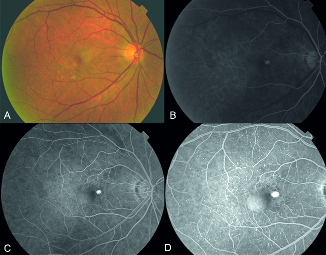
Figure 1.

A, Fundus photograph showing focal changes in the right eye at the macula, with the presence of drusen and focal thickening of the retina; B–D showing early to late phases of fluorescein angiography demonstrating hyperfluorescence in the area of focal thickening confirming the presence of a true classic CNV membrane associated with an adjacent sub-retinal pigment epithelial detachment
