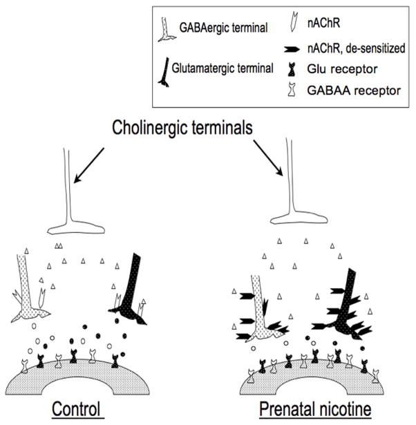
Figure 3.
Schematic diagram summarizing putative physiologic and anatomic changes that occur secondary to the influence of prenatal nicotine exposure on inhibitory synaptic transmission in brainstem respiratory neurons. Prenatal nicotine exposure results in an up-regulation of nicotinic acetylcholine receptors that are located presynaptically on both GABAergic and glutamatergic neurons. However, the nicotinic receptors are subsequently desensitized leading to a diminution of GABA and glutamate release. The reduction in GABA and glutamate release leads to an up-regulation of GABAA and glutamate receptors (probably both NMDA and AMPA subtypes) on the postsynaptic neuron. As a result, any endogenous stressor associated with an increase in GABA or glutamate release (e.g., hypoxia) would be associated with an exaggerated post-synaptic response (see text for detailed explanation). This model is an adaptation and extension of a similar one published by Luo et al. (Luo et al., 2007).

