
Figure 5.
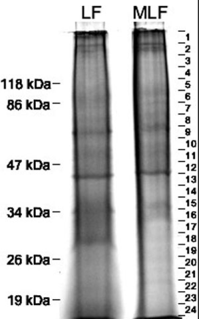
Electrophoresis of lipofuscin and melanolipofuscin proteins. Representative SDS-PAGE lanes of 50 μg of lipofuscin (LF) and melanolipofuscin (MLF) proteins. Mobility of molecular weight markers are indicated to the left. On the right, gel slices taken for subsequent in-gel digestion are shown. The lack of well-focused bands in the gel lane indicates microheterogeneous populations of the proteins, probably resulting from extensive modifications.