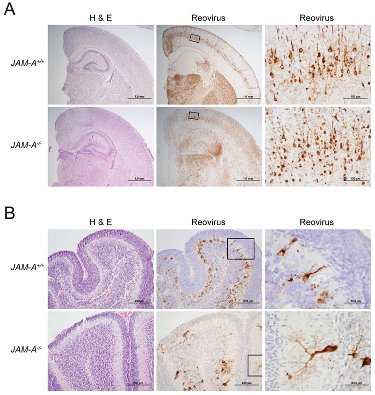
Figure 4. Histopathology of Reovirus Infection following Intracranial Inoculation.
Newborn JAM-A+/+ and JAM-A−/− mice were inoculated intracranially with 104 PFU T3SA-. Eight days after inoculation, brains of infected mice were resected and bisected sagittally. The left hemisphere was prepared for viral titer determination by plaque assay, and the right hemisphere was processed for histopathology. Consecutive coronal sections were stained with H&E or polyclonal reovirus antiserum. Representative sections of brain hemisphere, matched for hippocampal depth (A), and cerebellum (B) are shown. Boxes indicate areas of enlargement in the panels on the right and show cortical neurons (A) and cerebellar Purkinje neurons (B). JAM-A+/+ brain sections are from brains with left hemisphere viral titers of 4.1 × 10⁹ PFU (A) and 3.0 × 10⁹ PFU (B). JAM-A−/− brain sections are from brains with left hemisphere viral titers of 3.4 × 10⁹ PFU (A) and 1.6 × 10⁹ PFU (B).

