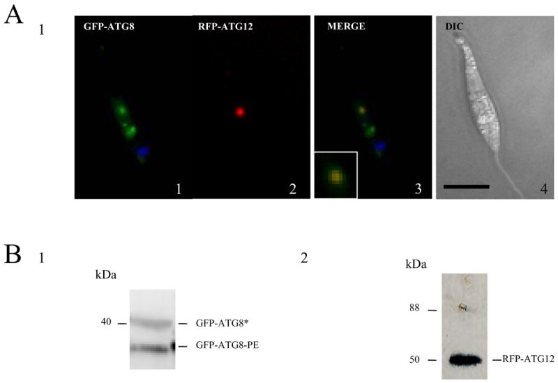
Figure 3. Localization of ATG12 in L. major promastigotes.

A. Occurrence of RFP-ATG12-containing structures (A2) and GFP-ATG8-containing putative autophagosomes (A1) in wild type L. major promastigotes expressing both proteins. The position of the kinetoplast is shown (blue) using DAPI staining. L. major procyclic promastigotes were suspended in nutrient-deprived medium for 120 min and observed by fluorescence microscopy. The merge of the RFP-ATG12 and GFP-ATG8 images, the co-labelled structure is enlarged at the bottom left, and the DIC image of the promastigote are shown in A3 and A4, respectively. Scale bar, 10 μm.
B. Western blot analysis using α-GFP (B1) and α-RFP (B2) antibodies on L. major procyclic promastigote cell extracts (5-8 × 10⁶ cells ml−1) transfected with GFP-ATG8 and RFP-ATG12. The α-GFP antibody detected a faster migrating lipidated ATG8 (labelled GFP-ATG8-PE) and a higher molecular mass unlipidated ATG8 (labelled GFP-ATG8*), resolved in a urea SDS-PAGE gel (B1). The anti-RFP antibody showed just a single protein (labelled RFP-ATG12) consistent with the predicted size of the fusion protein (B2).
