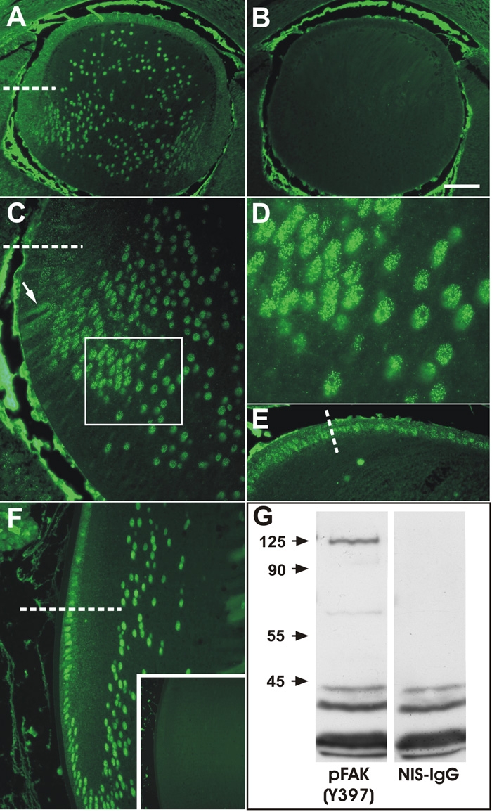
Figure 3.

Immunolocalization of phosphoY397-FAK. A: At E14.5 phosphorylated-FAK was detected as punctate reactivity throughout the anterior epithelium, associated predominantly with membranes and in the cytoplasm. In the transitional zone below the equator (dashed line) and in fiber cells, reactivity became increasingly associated with the fiber cell nuclei. B: Incubation with non-immune mouse IgG revealed no specific staining in the lens; non-specific reactivity is present in the aqueous and vitreous chambers. C: At E17.5 a similar pattern of reactivity was detected, with reactivity being predominantly associated with membranes of epithelial cells and early differentiating fibers (arrow) but becoming increasing punctate nuclear as fibers elongate and differentiate into fibers (box). D: High magnification view of boxed region in C, showing the distinct nuclear punctate reactivity. E: High magnification view of epithelial cells anterior to the equator (dashed line) showing predominantly cytoplasmic and occasional nuclear punctate reactivity. F: A similar pattern of reactivity was present at P21, with epithelial cells in the posterior germinative zone showing predominantly cytoplasmic reactivity and cells below the equator showing predominantly nuclear reactivity. G: Western blots of whole lens extracts showed a predominant specific band at the predicted 125 kDa molecular mass and a faint minor band at about 65 kDa. Incubation with non-immune IgG showed no specific bands and non-specific staining of low molecular weight complexes (<45 kDa). The scale bar in A, B, and F indicates 100 μm; in the inset 250 μm; in C, and F, 40 μm; and in D 16 μm.
