Fig. 3.
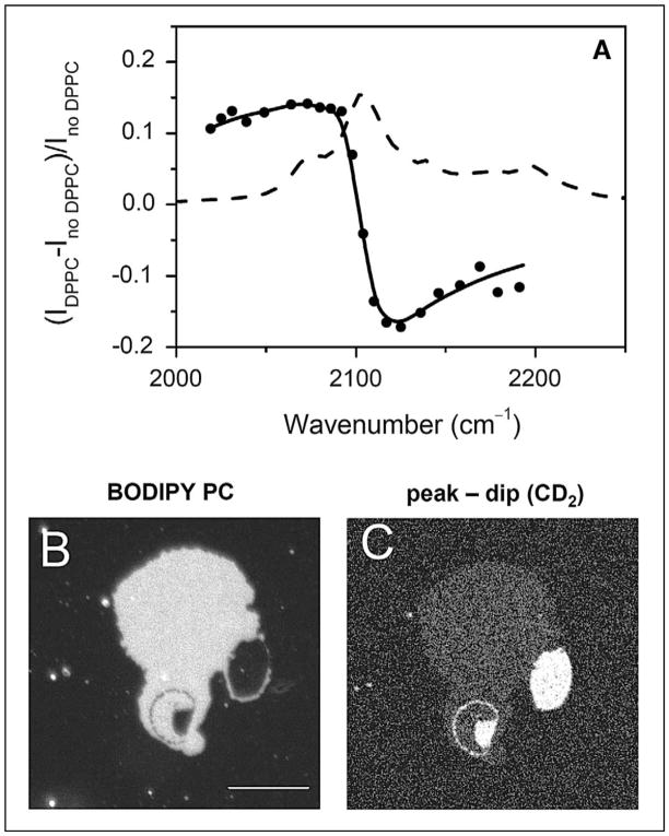
E-CARS imaging of lipid domains in a supported bilayer. (A) E-CARS spectral profile (solid line with solid circles) of a single supported bilayer of d62-DPPC in the C–D stretch vibration region. The peak and dip of the CARS band appeared at 2080 cm−1 and 2125 cm−1, respectively. The Raman spectrum (dashed line) of bulk d62-DPPC is also shown as a reference. (B) Fluorescence image of a DOPC/d62-DPPC (1:1) bilayer patch. Image was acquired at 23 °C. The liquid phase was labeled by BODIPY PC. (C) The (peak − dip) CARS image of the same sample, obtained by subtracting the CARS image at 2125 cm−1 from that at 2080 cm−1. The gel phase that is enriched in d62-DPPC exhibited a bright CARS contrast. Bar length = 10 μm. Data adapted from the paper by Li et al.53

