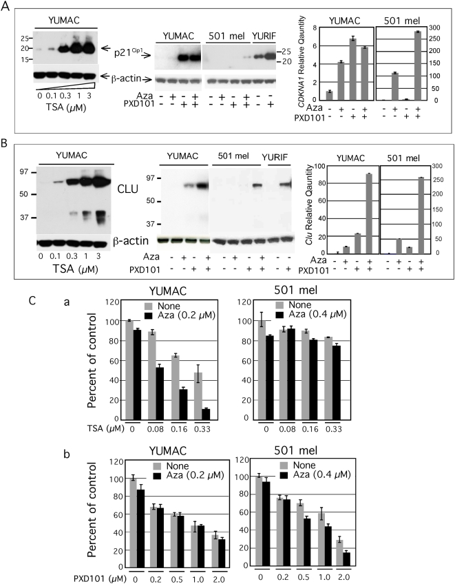
Figure 6. Reactivation of CDKNA1 and CLU by histone acetylation.
The Western blots show p21Cip1 (Panel A) and CLU (Panel B) expression in YUMAC melanoma cells treated with increasing concentrations of Trichostatin A (TSA) overnight, as revealed by probing with the respective antibodies using β-actin as a control (left panel). Middle panels show expression of p21Cip1 and CLU after 2-days treatment with Aza (0.2 µM), where indicated (+) followed by 1-day incubation with PXD101 (1 µm). Left panels: Real-Time PCR data comparing fold-difference in CDKN1A and CLU transcript in YUMAC and 501 mel cells after treatment with low-dose Aza and PXD101, alone and in combination, compared to non-treated cells. PXD101 (1 µM) was added for 24 hrs before harvesting the cells. Notice differences in scale of absolute hybridization intensities in YUMAC and 501 mel cells. However, 501 mel expressed about 370 fold less CDKN1A gene transcripts compared to YUMAC cells, apparently insufficient to lead to detectable p21Cip1 protein. The basal levels of CLU transcripts in YUMAC were about 50 fold higher relative to 501 mel melanoma cells, in agreement with low protein levels (data not shown). Panel C. Growth response to combination treatment with HDAC inhibitors Trichostatin A (TSA) and PXD101. The sensitive YUMAC and resistant 501 mel cells were incubated in triplicate wells without or with Aza (0.2 µM and 0.4 µM as indicated) followed by one-day recovery in regular medium, or medium supplemented with increasing concentrations of TSA (a), or PXD101 (b). Cell viability was assessed with the CellTiter-Glo® Luminescent Cell Viability Assay. Data are presented as percent of control, non-treated cells.

