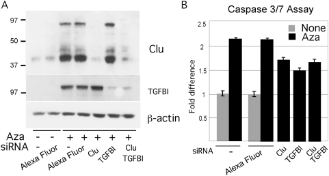
Figure 8. Validation of CLU and TGFBI apoptotic activity.
Panel A. Reduction of Clu and TGBFI proteins by gene specific siRNA knockdown assessed by Western blotting. YUMAC melanoma cells were untreated or treated with Aza (0.2 µM) for 2 days followed by transient transfection with siRNA directed to Clu, TGFBI, or a mixture of the two, employing Alexa Fluor as a control, as indicated. Cells were harvested the following day and extracts subjected to successive Western blotting with the respective antibodies, and anti-β-actin as a control. Panel B. Parallel cultures were tested for apoptosis with the Caspase 3/7 assay. Values are given as percent of control, i.e., non-transfected cultures.

