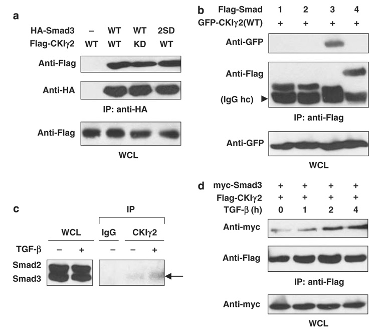
Figure 1. CKIγ2 constitutively and selectively interacts with Smad3.
(a) 293T cells were transfected with the indicated constructs. Cells were lysed in universal lysis buffer supplemented with protease and phosphatase inhibitors (ULB+) at 24 h post-transfection and anti-HA immunoprecipitation was performed. 2SD, S423/425D; KD, kinase-dead; WCL, whole-cell lysate. (b) The Flag-tagged, full-length Smad constructs were co-expressed with GFP-CKIγ2(WT). Anti-Flag immunoprecipitation was performed as in (a). The arrowhead indicates the heavy chain (hc) of the Flag antibody (M2). (c) Parental HaCaT cells were pretreated with 10 µM MG-132 for 1 h before treatment with or without 100 pm TGF-β for another 2 h. Endogenous CKIγ2 was precipitated with a goat polyclonal anti-CKIγ2 antibody (C-20), and endogenous Smad proteins were blotted with a monoclonal anti-Smad1/2/3 antibody. Co-precipitated Smad3 is indicated by an arrow. An irrelevant goat polyclonal antibody (IgG) was used as a negative control. (d) Wild-type Smad3 and CKIγ2 were co-expressed in MEFs for 20h. Cells were then treated with 100pm TGF-β for the indicated time course and then lysed for anti- Flag immunoprecipitation. CKIγ2, casein kinase 1 gamma 2; HA, hemagglutinin; MEFs, mouse embryonic fibroblasts; TGF-β, transforming growth factor-beta.

