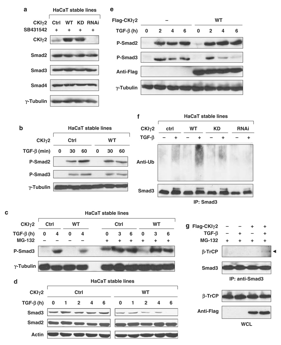
Figure 4. CKIγ2 promotes the proteasomal degradation of activated Smad3.
(a) CKIγ2 does not regulate the basal levels of Smad proteins. HaCaT stable lines were treated with SB431542 (10 µm) overnight, and endogenous Smad2, -3 and -4 were examined by western blotting. (b) The HaCaT lines were transiently stimulated with TGF-β (50pm) as indicated. C-terminal phosphorylation of endogenous Smad2 and Smad3 was measured. (c) HaCaT cells were treated with TGF-β (100 pm) for the indicated time courses in the absence (left) or presence (right) of 20 µm MG-132. The level of activated Smad3 was shown. (d) HaCaT cells were treated with TGF-β (100 pm) for the indicated time courses and the total levels of endogenous Smad2 and Smad3 were determined. (e) MEFs were transfected with or without wild-type CKIγ2 before incubation with TGF-β (50pm) for the indicated time course. C-terminal phosphorylation of endogenous Smad2 and Smad3 was measured. (f) HaCaT cells were pretreated with MG-132 (7.5 µM) for 1 h before the treatment of SB431542 (10 µm, ‘−’) or TGF-β (100 pm, ‘+’) for another 3 h. Cells were harvested in SDS lysis buffer (see Materials and methods), endogenous Smad3 was immunoprecipitated and ubiquitinated species of Smad3 was analysed by anti-ubiquitin blotting. (g) MEFs expressing either vector control or wild-type CKIγ2 were treated with SB431542 (10 mm, ‘−’) or TGF-β (100 pm, ‘+’) for 2 h in the presence of 20 µm MG-132. The interaction between endogenous Smad3 and β-TrCP was determined by co-immunoprecipitation assays. CKIγ2, casein kinase 1 gamma 2; MEFs, mouse embryonic fibroblasts; TGF-β, transforming growth factor-beta.

