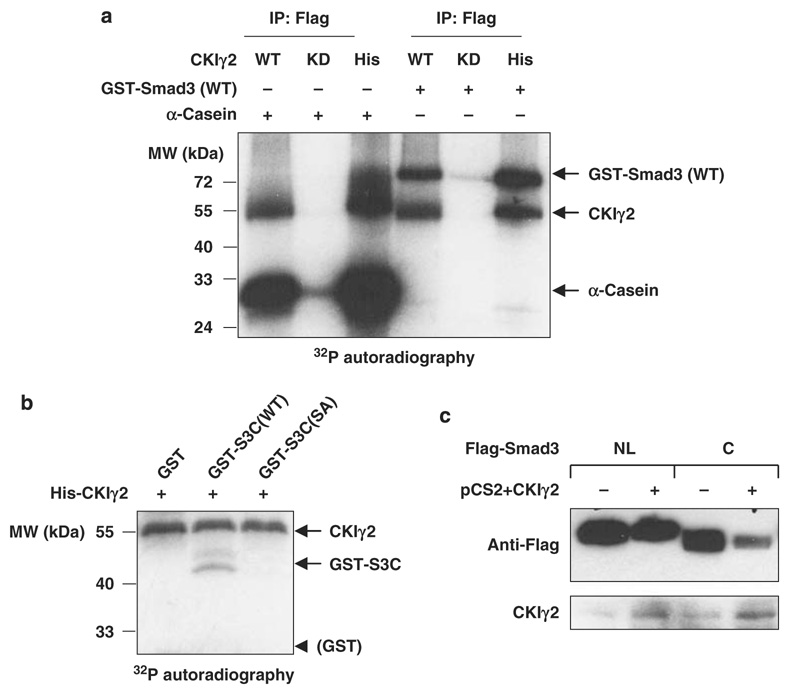
Figure 5. CKIγ2 phosphorylates Smad3 at Ser418.
(a) CKIγ2 in vitro kinase assay. Flag-tagged CKIγ2 (WT or KD) immunoprecipitated from 293T cell lysates as well as bacterially purified His-CKIγ2 were individually incubated with α-casein (positive control) or GST-Smad3(WT) in the presence of [32P]- γ-ATP. Phosphorylated proteins were visualized by autoradiography. Note that CKIγ2 underwent significant autophosphorylation. (b) His-CKIγ2 was incubated with GST alone, GST-S3C(WT) or GST-S3C( S418A) in a similar kinase assay as in (a). Equal loading of protein substrates was confirmed by Coomassie Blue staining (data not shown). CKIγ2 does not phosphorylate the GST moiety. (c) The indicated Flag-tagged Smad3 fragments were co-expressed with either a vector control or wild-type CKIγ2 in MEFs without inhibition of proteasomal function. Total cell lysates were analysed for the levels of Flag-S3NL (MH1 + Linker) and Flag-S3C (MH2). CKIγ2, casein kinase 1 gamma 2; GST, glutathione S-transferase; KD, kinase-dead; MEFs, mouse embryonic fibroblasts; WT, wild-type.

