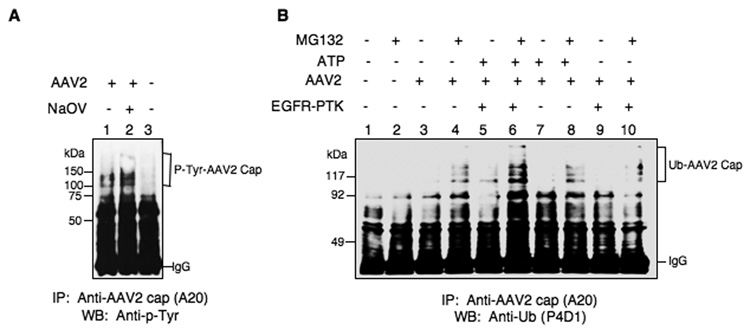
Fig. 5.
(A) Detection of intracellular tyrosine-phosphorylation of intact AAV2 vectors. Whole cell lysates (WCLs) prepared from HeLa cells untreated (lanes 1 and 3) or treated with NaOV (lane 2) following infection with K9 vectors (lanes 1 and 2) or mock-infection (lane 3), were immuno-precipitated with anti-AAV2 capsid antibody A20 followed by Western blot analyses with anti-p-Tyr antibody. (B) Detection of intracellular ubiquitin-conjugation of intact AAV2 vectors after in vitro phosphorylation of viral capsids by EGFR-PTK. Western blot analyses of ubiquitinated AAV2 capsid proteins in HeLa cells following transduction with ssAAV2-K9 vectors after in vitro phosphorylation of viral capsids by EGFR-PTK. Whole cell lysates (WCLs) prepared from HeLa cells untreated or treated with MG132 following mock-infected (lanes 1 and 2), or infected with K9 vectors, which were mock-incubated (lanes 3 and 4), or incubated with ATP (lanes 7 and lane 8), EGFR-PTK (lanes 9 and 10) or both (lanes 5 and 6), were immuno-precipitated with anti-AAV2 capsid antibody A20 followed by Western blot analyses with anti-Ub monoclonal antibody P4D1. This result is representative of two independent experiments.

