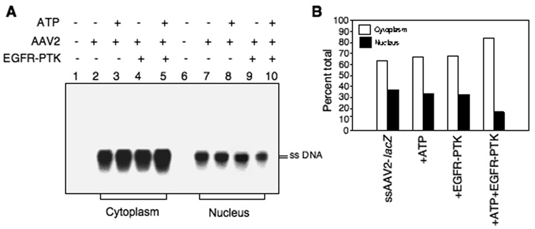
Fig. 6.
Detection of intracellular trafficking of AAV2 vectors to the nucleus after in vitro phosphorylation of viral capsids by EGFR-PTK. (A) Southern blot analyses of cytoplasmic and nuclear distribution of AAV2 genomes in HeLa cells after in vitro phosphorylation of AAV2 capsids by EGFR-PTK. HeLa cells were mock-infected (lanes 1 and 6) or infected by AAV2-lacZ vectors, which were mock-incubated (lanes 2 and 7) or pre-incubated with ATP (lanes 3 and 8), EGFR-TPK (lanes 4 and 9) or both (lanes 5 and 10). Nuclear and cytoplasmic fractions were prepared 18 hrs post-infection, low-Mr DNA samples were isolated and electrophoresed on 1% agarose gels followed analyzed by Southern blot hybridization using a 32P-labeled lacZ DNA probe. (B) Densitometric scanning of autoradiographs for the quantitation of relative amounts of viral genomes. These results are representative of two independent experiments.

