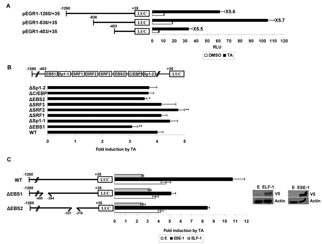
Figure 3. ETS binding site (EBS) located at −400 to −394 of the EGR-1 promoter is necessary for EGR-1 transcription induced by TA.
A Structures of EGR-1 sequential deletion constructs and deletion promoter assay. The EGR-1 promoter fragments of a different length but with the same 3'-end were cloned into pGL3-Basic. HCT-116 cells were co-transfected with 0.5 µg of each reporter construct containing the EGR-1 promoter and 0.05 µg of pRL-null vector using Lipofectamine. After growth overnight with fresh media, the cells were treated with 30 µmol/L of TA for 24 h. Luciferase activity was measured as a ratio of firefly luciferase signal/renilla luciferase signal and was shown as mean ± S.D. of three independent transfections. B The putative transcription binding sites within the −403 to +35 region in the EGR-1 promoter and luciferase assay with internal deletion clones. The boxes in the promoter represent the binding site of indicated transcription factors and are used for construction of internal deletion clones. HCT-116 cells were co-transfected with 0.5 µg of each internal deletion construct of the EGR-1 promoter and 0.05 µg of pRL-null vector using Lipofectamine and treated with 30 µmol/L of TA for 24 h. The X axis shows fold induction over vehicle as 1.0. The results are presented as means ± S.D. of three independent transfections. *, P < 0.05; **, P < 0.01 versus pEGR1-1260/+35 WT transfected cells. C Effect of ESE-1 overexpression on EGR-1 transactivation. HCT-116 cells were co-transfected with wild type EGR-1 promoter (pEGR1-1260/+35 WT or internal deletion clone pEGR1-1260/+35ΔEBS) in the presence of empty (E), ESE-1, or ELF-1 expression vector using Lipofectamine. After growth overnight with fresh media, the cells were treated with 30 µmol/L of TA for 24 h. The X axis shows fold induction over vehicle as 1.0. The results are presented as the means ± S.D. of three independent transfections. The overexpression of ELF-1 and ESE-1 was confirmed by Western analysis using V5 tag (GKPIPNPLLGLDST) antibody.

