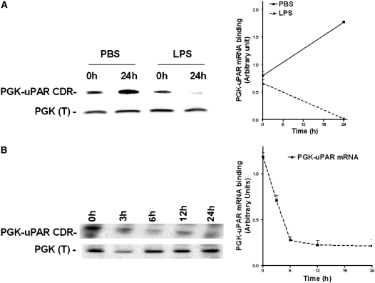
Figure 3.
Effect of LPS on the interaction of phosphoglycerate kinase (PGK) with urokinase-type plasminogen activator receptor (uPAR) mRNA coding region (CDR) in Beas2B cells. (A) Beas2B cells grown in culture plates to near confluence were incubated with phosphate-buffered saline or LPS (20 μg/ml) for 0 and 24 hours. PGK was isolated from these cells, as described in the methods, separated by sodium dodecyl sulfate (SDS)–polyacrylamide gel electrophoresis (PAGE), and transferred to nitrocellulose membranes. The membranes were then hybridized with 32P-labeled uPAR CDR mRNA. The same membranes were later stripped and analyzed for PGK protein by Western blot analysis using anti-PGK antibody. The line graph represents the ratio of uPAR CDR mRNA binding activity to PGK protein from three independent experiments shown as the mean ± SD. (B) Beas2B cells grown in culture plates to near confluence were incubated with LPS (20 μg/ml) for 0–24 hours. PGK was isolated from these cells, separated by SDS-PAGE and transferred to nitrocellulose membranes. The membranes were processed as described above. The line graph presents results from three independent experiments shown as the mean ± SD.

