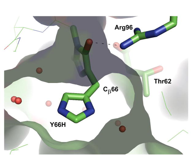
Figure 4.

Structural arrangement of Arg96 Nη2 in relation to Cβ66, as exemplified by the X-ray structure of the chemically reduced Y66H variant (10). The image was generated from pdb ID code 2fwq (10) using the program PyMol (36). The molecular surface of the chromophore binding pocket is also shown.
