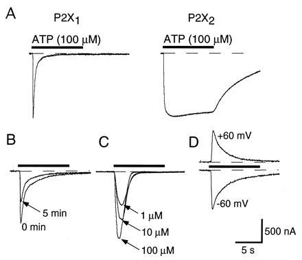
Figure 1.

P2X1 receptor desensitization. (A) ATP induces a rapidly desensitizing current when applied to oocytes expressing P2X1 receptors (Left) but a weakly desensitizing current in an oocyte expressing P2X2 receptors (Right). In this and subsequent figures, the ATP is applied for 10 s (indicated by horizontal bar above trace). Current traces have been normalized for comparison of time courses; the mean peak amplitude of currents at P2X1 receptors was 1600 ± 160 nA (n = 38) and at P2X2 receptors 3150 ± 260 nA (n = 47). (B) Desensitization at P2X1 receptors is unaffected by run-down of the response. The superimposed responses were evoked by 100 μM ATP, and applied for the first (0 min) and second times (5 min) to the same oocyte. The decline in the response is not associated with a change in the rate of desensitization. (C) Desensitization at P2X1 receptors is not strongly dependent on concentration. The superimposed traces were evoked by the concentrations indicated (10 s) applied at intervals of 20 min. (D) Desensitization at P2X1 receptors is not voltage-dependent. Two applications of 100 μM ATP for 10 s at an interval of 5 min are shown.
