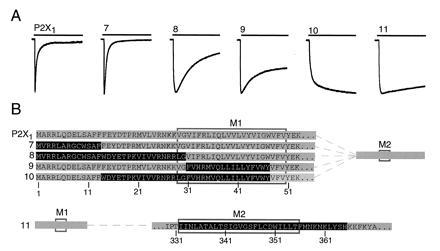
Figure 3.

Determination of P2X2 domains that prevent desensitization in the P2X1 receptor. (A) Representative currents evoked by 10-s applications of ATP (100 μM) to oocytes expressing six different chimeric receptors. (B) Schematic representation of chimeras: grey, P2X1; black, P2X2. Rectangles indicate positions of putative membrane-spanning regions M1 and M2. Upper five rows show P2X1 receptor and four chimeras in which N-terminal segments of P2X2 were inserted into P2X1. Lower row shows P2X1 receptor in which an extended second transmembrane domain of P2X2 receptor was inserted. Mean currents in chimeras 7-11 were (measured in nA, number of oocytes in parentheses) 440 ± 58 (3), 1130 ± 260 (14), 1190 ± 420 (4), 788 ± 487 (3), and 319 ± 56 (14), respectively.
