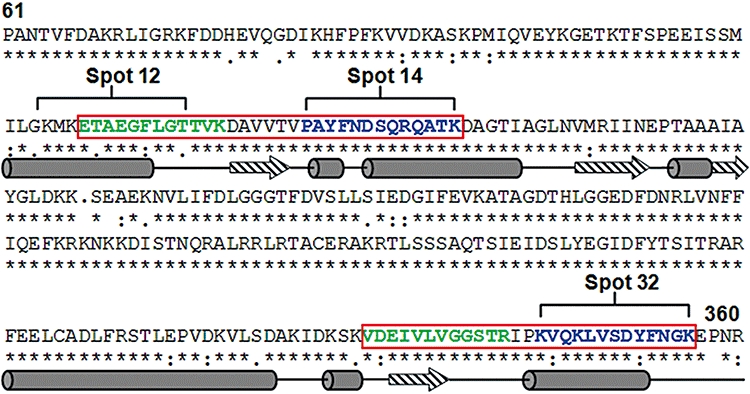
Fig. 5.

BHst 5-binding epitopes of Ssa2p are localized within the ATPase domain. The deduced primary sequences of Ssa1p and Ssa2p from C. albicans were aligned to show conserved regions (*) and variable regions (: or .). Hst 5 binding sites on Ssa2p identified by limited digestion (green) and peptide array (blue) (spots 12, 14 and 32) are indicated, and contiguous regions are enclosed by red boxes. Predicted secondary structure of BHst 5 binding regions was shown as α-helices (cylinders) and β-strands (arrows) below the primary sequences.
