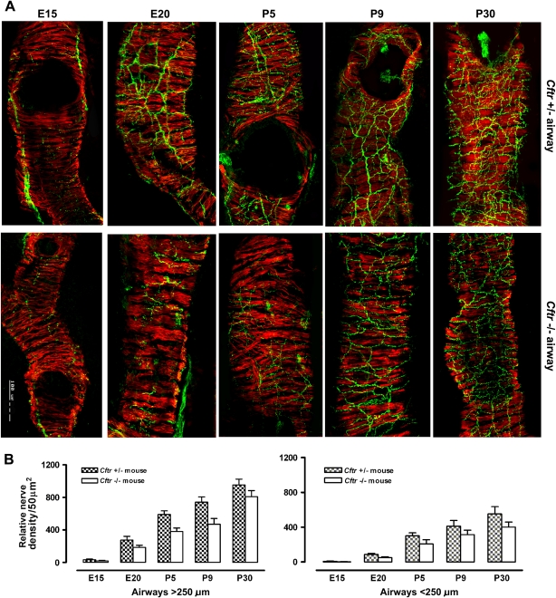
Figure 2.
(A) Composite photograph of confocal microscopy images of medium-size airways (100–200 μm) in the lungs of control (Cftr+/−) and Cftr null (Cftr−/−) mice during different developmental stages (E15, E20, P5, P9, P30) immunostained with SV2 and SMA antibodies. Well developed airway smooth muscle coat (red) is apparent even at an early (E15) developmental stage when only sparse nerve fibers (green) are noted. With progressing developmental stages, density and complexity of airway neural network increases in control but increases less in Cftr null mice. (B) Comparison of morphometric assessment of relative nerve density/50 μm2 within airway smooth muscle in different size airways in lungs of Cftr+/− versus Cftr−/− mice during development. Relative nerve density, expressed as means ± SD, in the airways of Cftr−/− mice is significantly reduced compared with Cftr+/− control mice (P < 0.001) in both airway sizes and during all developmental stages examined.

