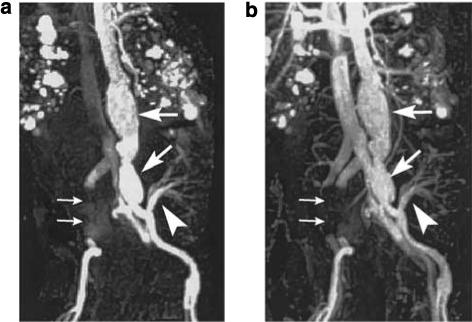
Figure 11. MR abdominal angiograms (TR/TE: 3.5/1.3 ms) of a 60-year-old male acquired after injection of fourfold diluted ferumoxytol (4 mg/Fe/kg).

(a) First-pass image. (b) Equilibrium image. Aortic and left iliac aneurysms (large arrows) are demonstrated. The artery arising from the left iliac artery (arrowhead) supplies a transplanted kidney. The right iliac artery and vein cannot be seen due to metal artifact from right iliac aneurysm stent (small arrows). Note that in the first-pass image, the inferior vena cava is visible. Polycystic kidney disease is also demonstrated.27 (First-pass contrast-enhanced magnetic resonance angiography in humans using ferumoxytol, a novel ultrasmall superparamagnetic iron oxide (USPIO)-based blood pool agent. (Reproduced from J Magn Reson Imaging 2005; 21: 46−52 with permission of Wiley-Liss, Inc., a subsidiary of Wiley Inc.)
